
What to notice
- Identify femur, tibia, fibula and patella before interpreting pathology.
- Differentiate articular cartilage from the medial and lateral menisci.
- Orient the ACL, PCL, MCL and LCL in relation to joint stability.
A clean 3D orientation view of the tibiofemoral and patellofemoral joints with the major stabilizing structures.
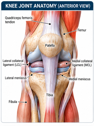
A clean 3D orientation view of the tibiofemoral and patellofemoral joints with the major stabilizing structures.
Detailed viewThe visual should be interpreted with the accompanying clinical context. PNA avoids using a single image or special test as stand-alone proof of a diagnosis.