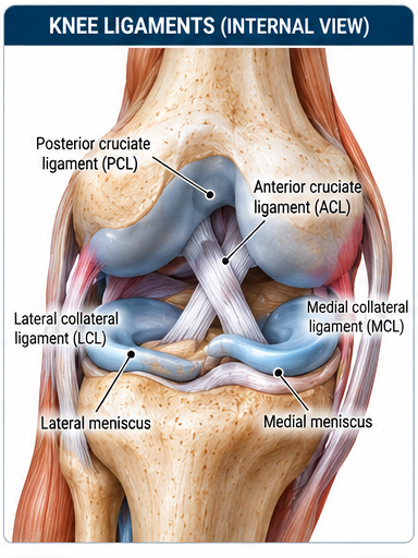
Partial or complete ACL disruption causing rotational and anterior knee instability, often during pivoting, deceleration or awkward landing.
THE MSK CLINICAL STORY
A pop, a pivot and the knee that lost trust in itself
A fictional teaching case. Follow the clues, enjoy the conversation, and make the decisions a thoughtful clinician would make—not an instant diagnosis from a single symptom.
Scene 01 · The patient walks in
A basketball player plants her foot, turns to pass and hears a pop. The knee swells quickly. By the next morning she says, 'The pain is calmer, so the ligament must be fixing itself, right?' The plot is not that simple.
A little clinic conversation
Patient: 'It's only painful when I try to turn.' Therapist: 'That's exactly why we care about the turning part.'
Scene 02 · Follow the clues
Pivot mechanism, rapid effusion and instability raise suspicion for ACL injury, often with associated meniscal or cartilage damage. Lachman findings add clinical evidence but are not a solo verdict.
Plot twist · What could fool us?
A better pain score does not prove restored rotational control. Nor does every ACL injury require the same surgical path; shared decisions depend on associated injury and goals.
Choose your learning lens
Same clinical case, two ways to understand it. The complete examination and treatment guide is still available below.
In everyday words
One of the knee's important stabilizing ligaments may be injured. We will check for associated damage, help the swelling and motion settle, and plan strengthening with the appropriate specialist if needed.
Scene 03 · The rehabilitation journey
Rebuild extension and quadriceps control first; then strength and single-leg mechanics; then running, deceleration, cutting and return-to-sport testing.
A pivot injury causes a pop, rapid swelling and a soft endpoint on Lachman. Which working hypothesis deserves priority?
Choose one answer to reveal the explanation. No score, no pressure—just better clinical reasoning.
These cases teach reasoning; they do not provide a diagnosis for an individual patient. Always consider red flags, contraindications, comorbidities and local referral pathways.
Possible associated meniscus, collateral ligament or osteochondral injury
Mechanism / Why It Happens
Non-contact pivot/cut with knee valgus and rotation
Sudden deceleration
Landing with poor control
Direct trauma in some cases
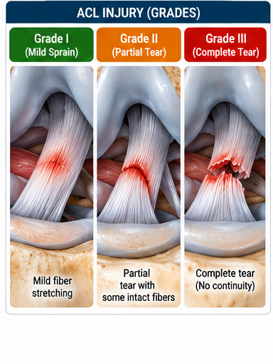
Classification / Grades
Grade I sprain
Grade II partial tear
Grade III complete tear; functional instability varies
Symptoms
Pop at injury
Rapid swelling/hemarthrosis
Giving way
Difficulty continuing sport
Pain and stiffness
Clinical Signs
Effusion
Loss of extension
Quadriceps inhibition
Anterior/rotational laxity
Altered gait
Red Flags & Referral Clues
Locked knee suggesting displaced meniscal tear
Neurovascular deficit
Suspected fracture/dislocation
Large acute effusion with severe pain requiring medical assessment
Diagnostic Examination
How to perform the tests — step by step
Each test includes patient setup, therapist position, hand placement, exact performance, positive finding and clinical interpretation.
Lachman Test
Assess ACL integrity.
Patient position
Supine; knee flexed about 20–30° with hamstrings relaxed.
Therapist position
Beside the tested knee.
Hand placement
One hand stabilizes the distal femur; the other grasps the proximal tibia.
How to perform
1Stabilize the femur.
2Translate the tibia anteriorly with a quick but controlled movement.
3Assess amount of translation and endpoint.
4Compare with the opposite knee.
Positive sign
Increased anterior translation with a soft or absent endpoint compared with the other side.
Interpretation
Strongly supports ACL injury when consistent with mechanism and history.
Anterior Drawer — Knee
Assess anterior tibial translation.
Patient position
Supine; hip about 45°, knee 90°, foot stabilized.
Therapist position
Sitting or standing near the foot.
Hand placement
Hands around proximal tibia with thumbs near the joint line.
How to perform
1Ensure hamstrings are relaxed.
2Stabilize the foot.
3Pull the tibia anteriorly.
4Compare side-to-side translation/end-feel.
Positive sign
Excess anterior tibial translation or soft endpoint.
Interpretation
Supports ACL insufficiency; can be less sensitive acutely due to guarding.
Pivot Shift
Assess functional rotational instability associated with ACL deficiency.
Patient position
Supine and relaxed.
Therapist position
At the tested leg.
Hand placement
One hand controls the heel/tibia; the other applies valgus force near the proximal tibia.
How to perform
1Start near extension.
2Apply internal tibial rotation and valgus stress.
3Slowly flex the knee.
4Observe/feel for tibial reduction around early flexion.
Positive sign
A palpable clunk/shift as the lateral tibial plateau reduces.
Interpretation
Supports ACL-deficient rotational instability; best performed by experienced clinicians.
Differential Diagnosis
Meniscal tear
MCL injury
Patellar dislocation
PCL injury
Osteochondral injury
Investigations / Imaging
MRI is commonly used to define ACL and associated injuries when indicated.
Radiographs may be required after trauma to exclude fracture.
Complete Rehabilitation Plan
From protection to full function
Phase 1 — Acute Recovery / Prehabilitation
Settle effusion, restore extension, recover quadriceps activation and normal gait.
What to do
Education and activity modification
Restore full extension early
Progress flexion
Quadriceps activation
Gait normalization
Shared orthopaedic decision-making for operative vs nonoperative pathway
Exercise dosage
Quad sets: 5–10 x 5–10 s, several times/day
Straight-leg raise if no extension lag: 3 x 8–15
Stationary bike when ROM permits: 10–20 min low intensity
Progress when
Trace/minimal effusion
Full extension
Improving flexion
No extension lag
Normalized walking
Phase 2 — Strength & Neuromuscular Control
Build quadriceps/hamstring strength and lower-limb control.
What to do
Progressive resistance training
Closed- and open-chain knee strengthening as appropriate
Hip/trunk strength
Balance and perturbation work
Exercise dosage
Squat/leg press: 3–4 x 6–12
Knee extension within appropriate tolerated range: 3 x 8–15
Hamstring strengthening: 3 x 8–15
Single-leg control drills: 3–4 sets
Progress when
Strength and movement quality improving
No reactive effusion
Good single-leg squat/step-down control
Phase 3 — Running, Plyometrics & Return
Restore high-speed force, landing, cutting and sport capacity.
What to do
Running progression
Jump/landing mechanics
Plyometrics
Change of direction
Sport-specific conditioning
Psychological readiness
Exercise dosage
Plyometrics: 2–3 sessions/week, graded contacts
Running volume/speed progressed independently
Agility exposure 2–3 sessions/week as tolerated
Progress when
No pain/effusion
Full ROM
Objective strength and hop symmetry appropriate for sport
Stable knee and satisfactory movement quality
Completed sport-specific progression
Manual Therapy — Role & Application
Patellofemoral/tibiofemoral mobilization may be used when joint mobility limits ROM.
Soft-tissue work can be an adjunct for short-term symptoms; it does not restore ligament stability.
Manual therapy should support, not replace, strength and neuromuscular rehabilitation.
Modalities — Use / Avoid
NMES may be useful when substantial quadriceps activation failure exists, especially post-operative/early rehab.
Cryotherapy/compression can help symptom and effusion management.
Passive modalities should not replace progressive exercise.
Return / Discharge Criteria
No pain or swelling
Full ROM
Stable clinical examination
High quadriceps/hamstring strength relative to sport demands
High-quality hop/jump/cutting performance
Psychological readiness
Full sport-specific training completed
Common Clinical Mistakes
Ignoring extension loss
Progressing despite recurrent effusion
Using time alone for return to sport
Neglecting quadriceps strength and landing/cutting mechanics
Clinical Pearls
A soft endpoint on Lachman after a pivot injury is a key ACL clue.
Rehabilitation quality matters whether the patient follows operative or nonoperative management.
Rapid substantial effusion after a pivot injury raises concern for significant intra-articular injury but is not specific to ACL rupture.
In an acutely painful, guarded knee, repeat examination after pain and swelling settle rather than forcing provocative tests.
Rehabilitation decisions should integrate instability episodes, activity goals, strength, movement quality and associated injuries—not MRI wording alone.
Before running, cutting or pivoting, assess progressive single-leg strength, landing control and sport-specific tolerance; time since injury alone is not clearance.
Continue learning
Connect assessment with management
Use the related PNA learning pathways to explore examination techniques and structured rehabilitation. A test result should always be interpreted alongside the history, full examination and clinical presentation.
Use these sources as a starting point for clinical learning. A reference being listed here does not, by itself, establish that every recommendation on this page is strongly supported. Check the original publication, study design, population and limitations before applying findings to an individual patient.
Educational resource only. Tests and treatment plans must be adapted to the individual patient, irritability, comorbidities, surgical restrictions and local scope-of-practice requirements.