Physio Nexus Academy logo
Physio Nexus Academy
MSK Injury & Rehabilitation Hub
ThighStrain / MuscleModerate evidence

Hamstring Strain

Musculotendinous hamstring injury commonly occurring during high-speed running or excessive lengthening.

THE MSK CLINICAL STORY

The sprinter who was fine at jogging—and not ready to sprint

A fictional teaching case. Follow the clues, enjoy the conversation, and make the decisions a thoughtful clinician would make—not an instant diagnosis from a single symptom.

Scene 01 · The patient walks in

A winger accelerates to chase a ball and feels a sharp snap in the back of the thigh. Weeks later he can jog and proudly announces, 'I'm cured.' His sport, however, has a second gear.

A little clinic conversation

Player: 'I ran slowly without pain!' Therapist: 'Excellent trailer. We still need to test the action scene.'

Scene 02 · Follow the clues

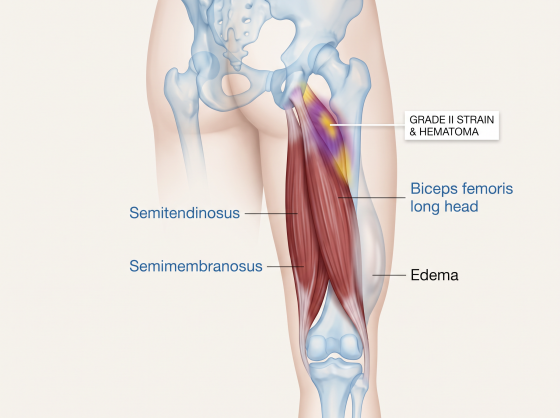
Sprint-type injury often involves the biceps femoris long head. Grade and location matter, and marked proximal weakness, bruising or a palpable defect may signal avulsion requiring referral.

Plot twist · What could fool us?

Overstretching a fresh tear can irritate it; never confuse early flexibility with restored high-speed force capacity.

Choose your learning lens

Same clinical case, two ways to understand it. The complete examination and treatment guide is still available below.

In everyday words

Some hamstring fibers may have been injured. Gentle, gradual strengthening comes before hard running, and return depends on what your sport actually asks your leg to do.

Scene 03 · The rehabilitation journey

Low-load contractions → strength at increasing muscle lengths → eccentrics and graded sprinting → sport-specific acceleration and braking.

See the full phased rehabilitation plan

You are the clinician · Interactive challenge

A footballer can jog pain-free after hamstring strain but has not done high-speed running. Best return-to-sport decision?

Choose one answer to reveal the explanation. No score, no pressure—just better clinical reasoning.

These cases teach reasoning; they do not provide a diagnosis for an individual patient. Always consider red flags, contraindications, comorbidities and local referral pathways.

PNA Clinical Visual Atlas

See this condition in 3D

Open the related anatomy and pathology visuals.

Open Visual Atlas

Structures Involved

  • Biceps femoris long head commonly in sprint-type injury
  • Semitendinosus
  • Semimembranosus
  • Proximal free tendon in some injuries

Mechanism / Why It Happens

  • High-speed sprinting
  • Sudden acceleration
  • Excessive hip flexion with knee extension
  • Kicking or overstretch injury

Classification / Grades

  • Grade I mild
  • Grade II partial tear
  • Grade III major/complete tear; proximal avulsion must be excluded

Symptoms

  • Sudden posterior-thigh pain
  • Pain with running
  • Pain on stretch or resisted contraction
  • Bruising in larger tears

Clinical Signs

  • Localized tenderness
  • Pain/weakness with resisted knee flexion or hip extension
  • Reduced tolerance to lengthened hamstring position
  • Possible palpable defect in severe injury

Red Flags & Referral Clues

  • Suspected proximal tendon avulsion
  • Marked weakness or palpable defect
  • Large hematoma
  • Neurologic symptoms suggesting lumbar/sciatic source
  • Inability to walk normally after severe injury

Diagnostic Examination

How to perform the tests — step by step

Each test includes patient setup, therapist position, hand placement, exact performance, positive finding and clinical interpretation.

Resisted Knee Flexion Test

Reproduce pain and assess hamstring force.

Patient position

Prone or sitting with the knee flexed to the chosen test angle.

Therapist position

At the tested leg.

Hand placement

Stabilize thigh/pelvis as needed; resistance applied at distal leg.

How to perform

  1. 1Ask the patient to flex the knee against resistance.
  2. 2Test at more than one knee angle when appropriate.
  3. 3Compare pain and force with the other side.

Positive sign

Concordant posterior-thigh pain and/or meaningful weakness.

Interpretation

Supports hamstring musculotendinous injury when localized to the injured region.

Passive Straight-Leg Raise / Length Test

Assess symptom response to hamstring lengthening.

Patient position

Supine.

Therapist position

Beside the tested leg.

Hand placement

Support heel/calf and maintain knee extension.

How to perform

  1. 1Keep knee extended.
  2. 2Passively flex the hip slowly.
  3. 3Stop at symptom onset or firm end-feel.
  4. 4Compare side-to-side and note symptom location.

Positive sign

Localized posterior-thigh pain or clearly reduced pain-limited range compared with the other side.

Interpretation

Supports length sensitivity; neural symptoms require separate neurodynamic interpretation.

Differential Diagnosis

  • Lumbar radiculopathy
  • Proximal hamstring tendinopathy
  • Adductor injury
  • Ischial apophyseal injury in adolescents
  • DVT when swelling/risk factors are concerning

Investigations / Imaging

  • MRI/ultrasound can help grade severe injury or suspected proximal tendon avulsion.
  • Imaging is not required for every mild strain.

Complete Rehabilitation Plan

From protection to full function

Phase 1 — Protect & Begin Loading

Settle acute irritability while preventing excessive deconditioning.

What to do

  • Avoid high-strain stretching/sprinting initially
  • Begin symptom-guided low-load contractions
  • Maintain hip/trunk and contralateral conditioning

Exercise dosage

  • Short-length isometric or low-load concentric knee flexion: 3–5 x 20–30 s or 3 x 10–15
  • Bridge variations: 3 x 8–12 if tolerated

Progress when

  • Normal walking
  • Low-load contraction tolerated
  • No increasing bruising/swelling

Phase 2 — Strength Through Range

Restore force across progressively longer muscle lengths.

What to do

  • Hip-dominant strengthening
  • Knee-flexor strengthening
  • Progress lengthened-state loading
  • Address lumbopelvic/kinetic-chain deficits

Exercise dosage

  • Romanian deadlift progression: 3–4 x 6–10
  • Slider/curl progression: 3 x 6–12
  • Split-stance hinge: 3 x 8–12

Progress when

  • Increasing outer-range strength
  • Minimal symptom response after loading
  • Jogging criteria met

Phase 3 — Eccentric, Running & Sprinting

Restore high-speed and sport-specific hamstring capacity.

What to do

  • Nordic/eccentric progression
  • Running exposure
  • Progressive sprinting
  • Sport-specific drills

Exercise dosage

  • Nordic: begin low volume such as 2 x 4–6 and progress
  • Running speed progressed in steps with pain-free sprinting as a late criterion
  • High-speed exposures separated by recovery days

Progress when

  • Near-full lengthened strength
  • Pain-free sprinting
  • Sport-specific high-speed demands tolerated

Manual Therapy — Role & Application

  • Gentle soft-tissue techniques can be used for comfort after the acute phase.
  • Avoid aggressive stretching or deep pressure over a fresh tear.
  • Manual therapy does not replace progressive loading and sprint re-exposure.

Modalities — Use / Avoid

  • Ice/compression may be used for short-term comfort in the acute phase.
  • Passive modalities have a limited role compared with progressive rehabilitation.

Return / Discharge Criteria

  • Pain-free high-speed running/sprinting
  • Restored outer-length and eccentric strength
  • Sport-specific acceleration/deceleration tolerated
  • No significant symptoms after previous loading

Common Clinical Mistakes

  • Aggressive stretching too early
  • Starting maximal eccentric loading immediately
  • Returning when jogging is pain-free but sprint exposure is absent
  • Ignoring kinetic-chain deficits

Clinical Pearls

  • Late rehab must include high-speed exposure for athletes who sprint.
  • Progress based on symptoms, strength and response to previous loading—not calendar time alone.

Continue learning

Connect assessment with management

Use the related PNA learning pathways to explore examination techniques and structured rehabilitation. A test result should always be interpreted alongside the history, full examination and clinical presentation.

Evidence sources

Use these sources as a starting point for clinical learning. A reference being listed here does not, by itself, establish that every recommendation on this page is strongly supported. Check the original publication, study design, population and limitations before applying findings to an individual patient.

Educational resource only. Tests and treatment plans must be adapted to the individual patient, irritability, comorbidities, surgical restrictions and local scope-of-practice requirements.