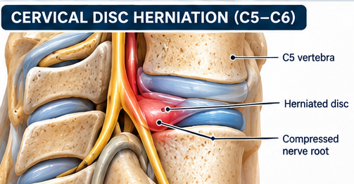
Cervical nerve-root disorder causing neck/arm pain with sensory, motor or reflex changes in a root distribution.
THE MSK CLINICAL STORY
The neck mystery that sent a message to the fingers
A fictional teaching case. Follow the clues, enjoy the conversation, and make the decisions a thoughtful clinician would make—not an instant diagnosis from a single symptom.
Scene 01 · The patient walks in
A programmer arrives rubbing the shoulder but points to tingling in the thumb and index finger. 'The trouble is in my neck. Why are my fingers getting notifications?' It is time to follow the nerve pathway.
A little clinic conversation
Patient: 'Is my arm wiring faulty?' Therapist: 'Maybe irritated, not necessarily damaged. We'll check the entire circuit.'
Scene 02 · Follow the clues
Neck-to-arm pain, dermatomal sensory changes, root-pattern weakness or reflex alteration may suggest cervical nerve-root involvement. Other peripheral nerve problems can imitate it.
Plot twist · What could fool us?
Tingling alone does not identify a root. Progressive weakness, gait disturbance or other myelopathy signs change the urgency of assessment.
Choose your learning lens
Same clinical case, two ways to understand it. The complete examination and treatment guide is still available below.
In everyday words
A nerve coming from the neck may be irritated, which can cause arm pain, tingling or weakness. We'll examine strength, sensation, reflexes and warning signs before deciding on treatment.
Scene 03 · The rehabilitation journey
First screen and calm symptoms → restore tolerable neck/upper-quarter movement → build strength, activity tolerance and a graded return to tasks.
A neck-pain patient develops progressive hand weakness and new gait imbalance. What should happen next?
Choose one answer to reveal the explanation. No score, no pressure—just better clinical reasoning.
These cases teach reasoning; they do not provide a diagnosis for an individual patient. Always consider red flags, contraindications, comorbidities and local referral pathways.
Repeated or directional movement when clearly beneficial
Gentle neural mobility
Monitor strength/reflex/sensation
Exercise dosage
Neck ROM/repeated movement: small sets of 5–10 based on directional response
Nerve sliders: 1–3 x 5–10, non-provocative
Progress when
Arm symptoms centralize/reduce
Neurologic status stable or improving
Phase 2 — Mobility & Strength
Restore cervical/scapular capacity and functional tolerance.
What to do
Deep neck flexor/endurance work
Scapular strengthening
Thoracic mobility
Progress neural mobility if indicated
Exercise dosage
Cervical endurance: 3–5 holds of 10–30 s
Rows/scapular strength: 3 x 8–15
General upper-quarter strength 2–3 days/week
Progress when
Work/ADL tolerance improving
Strength and sensation stable/improving
Phase 3 — Return to Demands
Restore work/sport lifting and sustained postures.
What to do
Progress resistance
Ergonomic/task conditioning
Overhead exposure if needed
Exercise dosage
2–3 progressive strength sessions/week
Progress when
No progressive neurologic deficit
Required tasks tolerated
Manual Therapy — Role & Application
Cervical/thoracic mobilization and traction may be used in selected patients as part of a multimodal program.
Avoid high-risk manipulation when vascular, instability, myelopathy or other contraindications are suspected.
Modalities — Use / Avoid
Traction may reduce symptoms in selected cases.
Heat/TENS may provide temporary comfort but should not replace neurologic monitoring and active rehabilitation.
Return / Discharge Criteria
Arm symptoms controlled
Neurologic findings stable/resolved
Functional strength and endurance restored
Work/sport postures tolerated
Common Clinical Mistakes
Calling any arm pain radiculopathy without neurologic exam
Provoking nerve tension aggressively
Missing progressive weakness or myelopathy
Clinical Pearls
Spurling may reproduce familiar arm symptoms and distraction may relieve them; neither finding alone establishes the diagnosis.
Progressive motor loss changes the urgency of management.
Record myotomes, dermatomes, reflexes and functional change at baseline and follow-up; a single provocative test should not establish the diagnosis.
Progressive weakness, signs of spinal cord involvement, gait/balance change, bilateral neurological symptoms or bowel/bladder disturbance require urgent medical assessment.
Use symptom response and neurological status to guide exercise and manual therapy; do not repeatedly provoke distal symptoms to demonstrate a positive test.
If symptoms or examination findings suggest peripheral nerve or shoulder pathology, revisit the differential rather than anchoring on a cervical source.
Continue learning
Connect assessment with management
Use the related PNA learning pathways to explore examination techniques and structured rehabilitation. A test result should always be interpreted alongside the history, full examination and clinical presentation.
Use these sources as a starting point for clinical learning. A reference being listed here does not, by itself, establish that every recommendation on this page is strongly supported. Check the original publication, study design, population and limitations before applying findings to an individual patient.
Educational resource only. Tests and treatment plans must be adapted to the individual patient, irritability, comorbidities, surgical restrictions and local scope-of-practice requirements.